Summary
Installed at the European synchrotron (ESRF) in Grenoble (BM32 beamline), the Laue microdiffraction instrument (µLaue) is unique in Europe and probes the matter by diffracting a polychromatic beam of a few hundred nanometers. The acquisition of the Laue diffraction diagram is very fast and allows scanning the samples to get the structural parameters of mono or polycrystalline materials in terms of orientation, crystallographic lattice parameters and state of deformations with a high precision. We added recently to this technique the possibility to record the emitted visible and near IR light excited from X-ray with the so-called XEOL technique (X-ray Excited Optical Luminescence). The acquisition of XEOL spectra (typically 1 s) can be easily synchronized to data collection of the Laue pattern so that to measure on the same specimen’s location. This new setup, just tested before the major upgrade of ESRF, has demonstrated its ability to correlate the crystal structural properties and the visible emission efficiency in nanoscale nitride heterostructures emitters (nanowires and µLEDs) and phosphors used for color management (i.e. white lightning). The master internship will consist in performing new ESRF experiments with the setup: improvement of light collection and setup as well as optimization of the data processing chains based on image analysis algorithms, intelligent Laue diagram recognition and micro and nanostructure reconstruction. Beamtimes will benefit from the Extremely Brilliant Source (EBS) upgrade of the ESRF to perform new experiments on µLEDs and nanowires. In parallel to ESRF activities, tabletop experiments will be performed with small external light sources, and full µLaue and XEOL datasets are already available to improve the data analysis procedure.
Full description of the subject
We propose to correlate the crystalline state properties and visible emission efficiency in nanoscale nitride heterostructures used in lightning emitters and µdisplays by mapping at the
same time µLaue and XEOL. This new (in the world) experimental development performed on BM32@ESRF
[0] combines the advantages of adapted beam size to get a full and fast information on
single objects, but also on
assemblies with a significant statistical information. It will provide unique information on growth achievements and integration in devices.
X-ray excited optical luminescence (XEOL) is an efficient technique to measure the optical properties of materials. A high X-ray flux combined with a high quantum efficiency of the materials is generally needed to get enough photons to collect the complete energy spectra of the optical emission. As an example, some original studies have been performed by our team at the (old) ID22@ESRF and (more recently) at the ID16B@ESRF beamlines with focused hard X-ray beam (size around 63x63 nm2, pink beam @ 29.6 keV & 17.5 keV). It allowed us to study the exciton confinement and radiative time decays for blue light emission in InGaN/GaN multiple quantum core-shell wells (MQW) deposited around GaN wires
[1], as well as the effect of crystalline polarity on Si-dopant incorporation and wire emission
[2]. These very small beams, providing a high spatial resolution, are well suited to study nanostructures, but mappings are quite long and only small area can be recorded (e.g. several hours of measurements per single dispersed wires on ID16). For these experiments, XEOL measurements have been combined advantageously with X-ray fluorescence (XRF) to correlate optical properties and indium composition inside multiple quantum wells (one of the most important parameters driving the emission color). It has been demonstrated that these wires have a very bright blue light emission and can be electrically excited. RGB color mixing is still not completely mastered and basic researches are still needed to understand device properties. Importantly, the determination of
strain and emission fluctuations
inside and
in-between devices is required. As an example, Ref.
[3] shows our realization of flexible white LED based on nitride wires and phosphors. These scientific questions in terms of
structural (strain) and optical fluctuations at the pitch scale and on the assemblies also apply in nitrides µ-Displays developed at CEA and III-V lab (cf L. Dupré, F. Templier’s team at Leti/DOPT). These structures are based on planar InGaN MQWs on c-planes and are electrically contacted to emit very bright light
[4].
During the internship, we will study (i) dispersed wires on nitride membranes and (ii) standing wires on sapphire with core-shell InGaN MQWs (diameter ~1.5 µm, length of the MQW ~10 µm) and (iii) 2D µDisplays with 6-7 µm pitch size. The samples will be selected from the MOVPE growths giving the best photo- and electro-luminescence performances. We demonstrated already on BM32 with similar wires the fast µLaue-scanning along the wires and the signature of MQWs periodicity and the setup has been significantly improved during the last years (new detector & higher stability of the beam). The light emitted by these materials is very bright (main wavelength in the 400-450 nm range) and can be recorded by the naked eyes. The XEOL setup collects the light with an off-axis parabolic Al-mirror (X-rays are going through the mirror), which is re-focused by means of a second parabolic mirror to the entrance of an optic fiber going to a spectrometer. The light is dispersed by a diffraction grating on a back-illuminated pixels CCD camera cooled by Peltier effect. µLaue analysis will be performed by the LaueTools program developed by J.S. Micha at the beamline to determine the strain for the full set of patterns of the mapping
[5,6]. For the XEOL signal, we are developing a specific python routine to analyze hyperspectral data. All the measurements will be done with line-scans and grid mappings with 0.3-0.4 µm beam size and will correspond to a very large quantity of data that can be treated by ESRF servers and optimized codes.
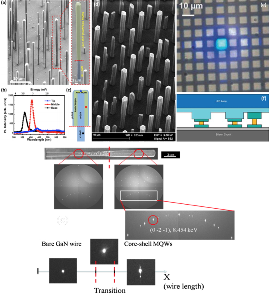
Top: Samples to be studied: (a) self-organized wires that will be dispersed on glass, (d) standing wires grown on sapphire. (c) shows the MQW heterostructures and (d) an example of photoluminescence spectra. (e) is a µDisplay structure with its technological integration shown in (f).
Bottom: Evolution of the scattering patterns collected by Laue Microdiffraction as a function of beam location along the nanowire. At the wire tip (right hand side) the multiple components shape of Laue peaks correspond to the periodic structure of the MQWs.
Requested skills
Strong interest in materials science and structural & optical properties as well as python programming for data analysis.
Keywords
synchrotron, µLaue, XEOL, LED, phosphors
ContactJean-Sébastien Micha,
DRF/IRIG/DIESE/SyMMES/STEP ESRF, The European Synchrotron, CRG-IF, ligne BM32, 71 Avenue des Martyrs, 38000 Grenoble
Phone number: +33 4 76 88 25 89
Master/Thesis supervisorJoël Eymery, CEA/IRIG/DEPHI/MEM/NRS, NRS
lab publications Head of CEA "Nanostructures and Synchrotron Radiation” Laboratory
Univ. Grenoble Alpes, IRIG, MEM, NRS, 17 rue des Martyrs, 38054 Grenoble, France.
Phone number: +33 4 38 78 30 15, joel.eymery@cea.fr