Summary
Installed at the European synchrotron (ESRF) in Grenoble (BM32 beamline), the Laue microdiffraction instrument (μLaue) is unique in Europe and probes the matter by diffracting a polychromatic beam of a few hundred nanometers. The acquisition of the Laue diffraction diagram is very fast and allows scanning the samples with a high precision to get the structural parameters of mono or polycrystalline materials in terms of orientation, crystallographic lattice parameters and state of deformations. We added recently to this technique the possibility to record the emitted visible and near IR light excited from X-ray; the so-called XEOL technique (X-ray Excited Optical Luminescence). The acquisition of XEOL spectra (typically 1 s) can be easily synchronized to data collection of the Laue pattern so that to measure on the same specimen’s location. The PhD subject consists in participating to the development of the analysis of new ESRF experiments with our team (improvement of light collection and setup) and more specifically to the optimization of the data processing chain based on image analysis algorithms, intelligent Laue diagram recognition, and micro and nanostructure reconstruction. This new data treatment will allow the systematic treatment of a large amount of data corresponding to objects that can be also randomly oriented. This “serial crystallography” (term used in biology) will be also correlated to the optical properties of important optoelectronic materials such as nitride materials. This work will benefit from the Extremely Brilliant Source (EBS) upgrade of the ESRF, recent fast pixel detector and continuous development of the LaueTools program for diffraction pattern analysis.
Full description of the subject
Context and in-house development: Nitride heterostructures demonstrated novel optical and electronic properties making use of quantum confinement effects and strain engineering (see the 2014 Nobel prize for blue nitrides LEDs). The emergence of disruptive functionalities is strongly related to the growth and technology controls, but also to the development of advanced characterization techniques having high spatial and spectral resolution. Focused X-ray beams provide innovative solutions to analyse quantitatively the morphology, defects, strain and composition of these materials.
This PhD project proposes to correlate the crystalline state properties and visible emission efficiency in nanoscale nitride heterostructures used in lightning emitters and μdisplays by mapping at the same time μLaue and XEOL. This new (in the world) experimental development performed on BM32@ESRF
[0] combines the advantages of adapted beam size to get a full and fast information on
single objects, but also on assemblies with a significant statistical information. It will provide unique information on growth achievements and integration in devices.
The X-ray excited optical luminescence (XEOL) is an efficient technique to measure the optical properties of materials. A high X-ray flux combined with a high quantum efficiency of the materials is needed to get enough photons to collect the complete energy spectra of the optical emission. As an example, some original studies have been performed by our team at the ID22 and ID16B@ESRF beamlines with nano-focused hard X-ray beam (size around 63x63 nm2, pink beam @ 29.6 keV & 17.5 keV). We studied the exciton confinement and radiative time decays for blue light emission in InGaN/GaN multiple quantum core-shell wells (MQW) deposited around GaN wires
[1], as well as the effect of crystalline polarity on Si-dopant incorporation and wire emission
[2]. These very small beams providing a high spatial resolution are well suited to study nanostructures, but mappings are quite long and only small area can be recorded (e.g. several hours of measurements per single dispersed wires on ID16). For these experiments, XEOL measurements have been combined advantageously with X-ray fluorescence (XRF) to correlate optical properties and indium composition inside multiple quantum wells (one of the most important parameters driving the emission color). It has been demonstrated that these wires have a very bright blue light emission and can be electrically excited. RGB color mixing is still not completely mastered and basic researches are still needed to understand device properties. Importantly, the measurements of
strain and emission fluctuations inside and
in-between devices are required. As an example, Ref.
[3] shows our realization of flexible white LED based on nitride wires and phosphors. These scientific questions in terms of
structural (strain) and optical fluctuations at the pitch scale and on the assemblies also apply in nitrides μ-Displays developed at CEA and in new screens. These structures are based on planar InGaN MQWs on c-planes and are electrically contacted to emit very bright light [4] with a main emission wavelength in the 400-450 nm range. The μLaue/XEOL setup has been significantly improved during the last year (new detector & higher stability of the beam). The XEOL is collected by an off-axis parabolic Al-mirror (X-rays are going through the mirror), which is re-focused by means of a second parabolic mirror to the entrance of an optic fiber going to a spectrometer. The light is dispersed by a diffraction grating on a back-illuminated pixels CCD camera cooled by Peltier effect (see Fig. 1). μm beams will be used to tackle the physical questions related to opto-electronic devices, to provide statistical information on a large number of emitters and estimate their fluctuations related to growth and technology process defects. A new analysis method has to be implemented that must combine high-level experiments experimental and data analysis.
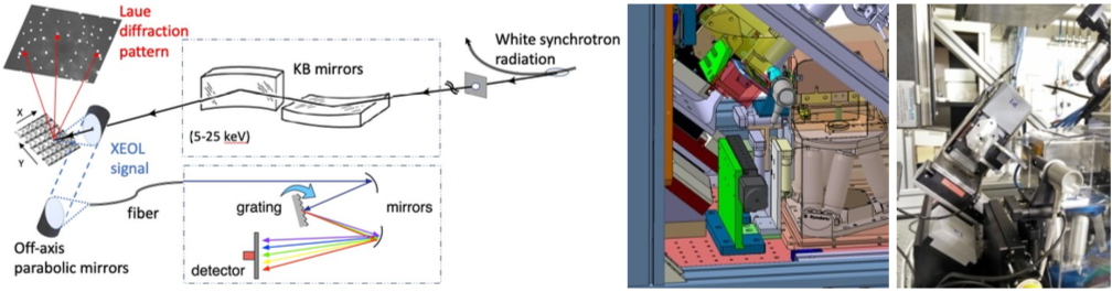
Fig. 1:
Left: Schematics of the experimental setup.
Right: drawings and picture of the μLaue setup of the BM32 beamline at ESRF.
Objectives: At the beginning of the PhD, the experimental work performed with the BM32 staff will be focused on (i) dispersed rods/wires on nitride membranes and (ii) standing wires on sapphire with core-shell InGaN MQWs (diameter ~ µ1.5 μm, length of the MQW ~ µ10 μm) and (iii) μDisplays grids with μm pitch size. The samples will be selected from the MOVPE growths (in-house research of the lab.) giving the best photo- and electro-luminescence performances suitable for optimized devices. The high-resolution fast mapping of the crystal properties with X- rays is a big challenge. By using polychromatic wavelength, the sample (see Fig. 2) does not need to be aligned to record the diffraction pattern and it needs only to be scanned.
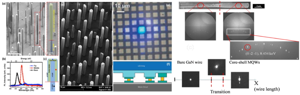
Fig. 2: Left: Samples to be studied: (a) self-organized wires that will be dispersed on glass, (d) standing wires grown on sapphire. (c) shows the MQW heterostructures and (d) an example of photoluminescence spectra. (e) is a μDisplay structure with its technological integration shown in (f). Right: Evolution of the scattering patterns collected by Laue Microdiffraction as a function of beam location along the nanowire. At the wire tip (right hand side) the multiple components shape of Laue peaks correspond to the periodic structure of the MQWs.
As already said, this technique is unique in Europe and only two similar instruments exist in the US and in Taiwan. It will soon benefit from the major upgrade of the ESRF synchrotron: the brilliance will be increased by a factor of 100 and beam size will be decreased, urging the necessity to speed-up the acquisition and analysis treatment to increase the throughput and the scientific & technological impact of this instrument. In this work, a recent 4096x4096 pixels X-ray SCMOS seamless camera array with 0.072 s readout time (Photonic Science) will be used. The data size becomes significant: a small map of 200x200 positions (for example with a probe size of 0.5 μm and a counting time of 01. s/point) generates about 320 Go of data (40000 images of diffraction pattern of about 8 Mo). We planned also in the next years to extend the μLaue technique to the completely new energy-dispersive pnCCD technology: a CDD pixel will then be encoded in X-ray energy... In the time scale, we also expect to operate the system at 400 Hz frame rate, still maintaining the full spectroscopic performance with a simultaneous measurement of diffraction. Presently, the μLaue analysis is performed by the LaueTools program developed by J.S. Micha at the beamline to determine the orientation and strain for the full set of patterns of the mapping
[5, 6]. For the XEOL signal that is less heavy (1024 wavelength channels), we are developing a specific python routine to analyze hyperspectral data.
The candidate will first interact with experimentalists to design better experiments, e. g. compressed sensing to optimize data collection with respect to a given physical property (for example statistical info on strain or light emission in heterogeneous samples). This work will also participate to the development of metadata treatment on large scale facilities,
e.g. the
Datahub /
ICAT+ and
PaNOSC (photon and neutron open science cloud) initiatives of the ESRF.
The improvement of the data analysis itself will be performed at three levels to get:
(i)
faster data processing - to improve specific points and performances of the LaueTools program and to develop parallel computing architectures (either massively many-core or GPU-based).
(ii)
better processing - to implement recent data treatments. First the goal will be to be able to quickly assign each peak to its parent crystal, in each image of a large image series and then to find the corresponding orientation. Such fast analysis is necessary because the experiments are conducted in limited timeframe. Any feedback on the data quality (
e.g., how interesting is the region being mapped? is the signal-to-noise ratio sufficient to distinguish different components? ...) is highly valuable to help in driving the experiment. To that end two approaches can be evaluated, using either supervised or unsupervised machine learning.
- Supervised: the peaks of the Laue pattern of a grain are linked by symmetry relations and thus can be classified by pattern matching approaches
[7].
- Unsupervised: using an auto-encoder on the detector images, the series can be analyzed in terms of Principal Components to identify the different grains
[8].
(iii)
better interpretation for multiple datasets -
e.g. Automation scripts. Principal Component Analysis (PCA and variants) to perform exploratory data analysis for making predictive models. Hyperspectral treatment of XEOL data,
e.g. with an
HyperSpy extension.
The candidate will benefit from the interactions with data analysis specialists at ESRF (see
PaNdata ESRF contributions) and French-CRG in the big data challenge, but also from the general efforts of CEA (Grenoble platform involved in Data Analysis).
Expected impact and perspectives: Large scale facilities are facing recently the “data deluge” due to the continuous development of new techniques with powerful excitation sources, improved sample and sensor positioning, faster detectors with a strong increase in size and number of pixels. The digitization of these tools has also been a revolution which is still ongoing. It will contribute actively to the materials characterization at the nanoscale, often mandatory for many process and device developments. This PhD will provide the missing element to reduce considerably the operation time of the μLaue/XEOL nano-characterisation which has the capability to improve the physical understanding of the growth and to control the quality of complex nanomaterials and opto-electronic devices. A new methodology, which will combine different analysis techniques of nano-characterisation and new computational tools is proposed. It corresponds to a new and necessary improvement of the nanomaterial’s characterization chain.
In addition to addressing important materials for applications (in Europe the Aledia, Plessey, Glö companies are developing actively nitrides μLEDs), the data analysis developed for these techniques will benefit to academic studies, to the synchrotron community (the French Collaborative Research Group, the ESRF and more generally Large-Scale infrastructures) and can be easily extended to many other topics.
The key objective of this project is to foster the collaborations between experimenters developing advanced characterization techniques and computer scientists, bringing their necessary skills in data analysis.
Candidate profile:
Requested skills: A master in data analysis or a master of Science (physics or nanoscience disciplines with strong motivations in data analysis) is required. Skills in machine learning, image analysis and Python/Notebooks (programming language at ESRF) are important. Preliminary experience in software development for parallel computing architectures (either massively many-core or GPU-based) is welcome. Curiosity, enthusiasms for physics, experiments as well as materials science will be appreciated.
Location: The PhD candidate will join the Nanostructure Radiation Laboratory (NRS) of the CEA including research scientists in physics and instrumentation. NRS contributes to the scientific and technological objectives defined by CEA and by the French community within the framework of the French X-ray synchrotron large scale facilities. These tasks require very specific synchrotron equipments and scientific skills to carry out high-level experiments on a broad range of subjects supporting both academic research and societally relevant applications. He/She should demonstrate excellent oral and written communication skills in English. Application: Please submit electronically (in PDF format) a detailed resume and a motivation letter.
Applications must be sent to Dr.
Joël Eymery, Dr
Samuel Tardif and Dr
Jean-Sébastien Micha. In the object of your application email, please include the reference “[PhDThesis-Application]” followed by your NAME and Surname.
Solid state Physics, Materials Physics
Ecoles Doctorales
Ecole doctorale de Physique de Grenoble
Contact person
Jean-Sébastien Micha, CEA/IRIG/DIESE/SyMMES/STEP,
ESRF, The European Synchrotron, CRG-IF, BM32 beamline, 71 Avenue des Martyrs, 38000 Grenoble
Phone number: +33 4 76 88 25 89
"Nanostructures and Synchrotron Radiation” Laboratory
Univ. Grenoble Alpes, IRIG, MEM, NRS, 17 Avenue des Martyrs, 38054 Grenoble, France.
Phone number: +33 4 76 88 28 19
Thesis supervisor
Joël Eymery, CEA/IRIG/DEPHI/MEM/NR,
NRS lab publications
Head of CEA "Nanostructures and Synchrotron Radiation” Laboratory
Univ. Grenoble Alpes, IRIG, MEM, NRS, 17 rue des Martyrs, 38054 Grenoble, France.
Phone number: +33 4 38 78 30 15