XRF microscopy is an analytical technique that uses a focused X-ray beam combined with the detection of fluorescent X-rays emitted by excited atoms. By scanning the sample point by point, it provides quantitative maps of chemical elements with a resolution that can reach the nanometer scale; however, this requires long acquisition times and high radiation doses, which can be critical for radiation sensitive samples such as biological cells or
energy materials*. Multichannel detectors, offer a trade-off between data quality and sample preservation by collecting more photons and increasing the
signal-to-noise
ratio (SNR). However, the number of detectors that can be integrated is limited by their cost and available space. An alternative is to attempt to improve the SNR using computational noise reduction methods.
In this study, researchers developed a self-supervised denoising based on the
Noise2Noisemethod* and the
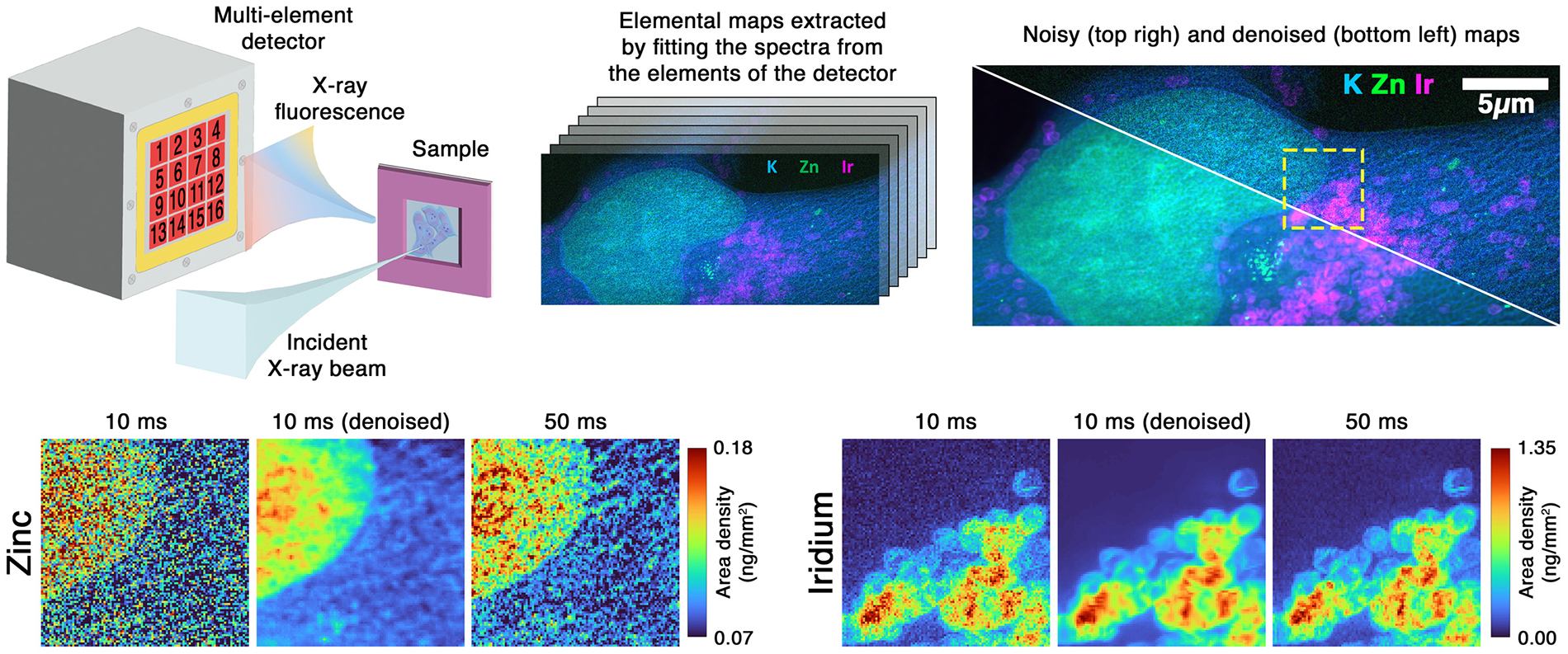
U-Net architecture*. This method was initially tested on a sample with a known geometry (“Siemens star*") and demonstrated a significant noise reduction in image without altering the geometry of the patterns or the quantification, even for a multi-element detector with only a few channels (2 to 4). The researchers subsequently validated this method on human cancer cells—a case representative of biological applications where it is imperative that radiation doses remain low—by showing that, based on fast acquisitions (10 ms per pixel), the resulting images, which were initially very noisy, are more readable, revealing structures that were obscured in the raw images, and are sharper than reference images acquired with five times longer exposure (50 ms per pixel).
© CEA-Irig/MEM
Figure: Diagram of the multi-element XRF detector arrangement alongside the denoising results. The panels compare raw data acquired at 10 ms per pixel, the denoised reconstruction, and a reference map recorded at 50 ms per pixel for Iridium and Zinc elemental lines.
The developed method enables obtaining images of comparable or even superior quality to those obtained through much longer exposure times, thus keeping the radiation dose low and acquisition time fast. This approach will therefore be particularly useful for mapping trace elements in radiation-sensitive biological samples, for characterizing materials requiring large-area analysis, and for time-resolved studies where acquisition speed is critical. Furthermore, to achieve this type of result, there is no need to use detectors composed of large number of detector elements, which means that this method can be implemented on existing XRF and EDX instruments without any hardware modifications.
energy materials*: materials used to generate, convert, store, or conserve energy, such as for manufacturing battery electrodes and electrolytes or produce, store, or use hydrogen. They encompass also thin film photovoltaic materials or materials that can convert CO₂ into useful molecules, …
Noise2Noise method*: a deep learning method that trains a denoising network using noisy images. Since noise is random and varies from one image to another, the network does not learn to reproduce the noise but gradually learns to identify the common element: the actual signal.
U-Net architecture*: a
convolutional neural network* architecture originally developed for image segmentation and now widely used for image restoration tasks. Its structure allows the network to analyze an image at multiple scales and reconstruct it while preserving fine details.
convolutional neural network*: a deep learning architecture that, through convolution operations, learns to recognize relevant structures in an image, thereby enabling denoising, segmentation, or restoration.
Noise2Noise method/U-Net-type architecture*: by treating the maps from the detector’s individual channels as independent observations of the same underlying structure, pairs of images are generated and used to train a denoising model. A denoised image is then reconstructed by combining the outputs of the trained U-Net network.
Siemens star*: a resolution test target made of radial gold patterns deposited on a silicon substrate.
UMR : MEM/LEMMA & MEM/NRX : CEA, Univ. Grenoble Alpes (UGA).
Fundings: ESRF beamtime at ID16A (experiment IHLS-3625); EU Horizon 2020 MSCA COFUND ENGAGE (No. 101034267) & SCANnTREAT (No. 899549).
Collaborations: ESRF.