The principle of ESR is simple: the studied sample is placed in a static magnetic field that orientates the electronic spins. These are then manipulated and detected by pulses of very short and very intense hyperfrequency waves (a few GHz). Because ESR detects magnetic moments associated with electronic spins, it is possible to study all the compounds bearing unpaired electrons (free radicals, metal ions in specific oxidation states, triplet states, ferromagnetic systems …). In contrast, ESR is completely blind to the diamagnetic systems that constitute the vast majority of the matter that surrounds us. At first glance, it may be seen as a severe limitation. Actually, this is a major advantage because a variety of small paramagnetic sub-systems are created from diamagnetic systems in response to perturbations (ionizing radiations, photons, oxidation, ageing processes...). ESR can thus study just the active parts in the middle of a sea of diamagnetic systems: it seems like the microscopic version of the search for a needle (paramagnetic) in a haystack (diamagnetic).
Let’s take the example of a photovoltaic device: light interacts with the components (pigments, doping, silicon…) that are initially diamagnetic, and creates excitons, triplet states, pairs of radicals… all paramagnetic. We study these phenomena on a model system, for example a photoactive compound like C60, directly irradiated in an ESR spectrometer, usually with a pulsed laser. Using specific detection methods, it is possible to analyze the paramagnetic species present in the sample as a function of time with a resolution of a few nanoseconds. The result is bidimensional maps (Figure 1). In our laboratory, over the years we have developed a pulsed ESR coupled to a laser. More recently, we have purchased an optic device allowing us to generate laser pulses of a duration of a few nanoseconds on a continuous range of wavelengths (0.4 – 2 µm). It is thus possible to study physical phenomena involved in these systems as a function of the wavelength.
Do we have a totally efficient spectroscopic method? Almost! In order to accurately study individual systems (for instance in nanostructures), the sensitivity of the approach is sometimes too low. How could we increase it? By optical detection or detection of electrical current. In this case, the electromagnetic wave of the ESP spectrometer is used to manipulate the electronic spins of the photoinduced species in order to modify their yield of recombination or dissociation. This results in a change of the fluorescence or photocurrent intensity directly and very efficiently detectable by the pODMR or pEDMR methods (Optically or Electrically Detected Magnetic Resonance) (Figure 2). They combine a sensitivity increase of a factor of 106 compared with classical ESR with all the possibilities of pulsed ESR spectroscopy. This apparatus is under development in our laboratory. It will be unique in France and comparable with the best systems designed in Germany and in the United States. It will be used in particular for our research on new technologies for energy and on quantum calculation.
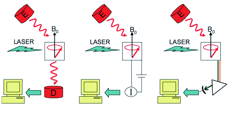
Figure 2: In a classical spectrometer (left), the manipulation of spins and the detection are done with microwaves (in red). In an EDMR system (middle), the manipulation of spins is still done with microwaves but the detection relies on the measurement of a photocurrent. Last, in the case of an ODMR apparatus, the detection is done through a monochromator and a photomultiplier which collect the photons emitted by the sample.