You are here : Home > LEMMA team > Projects and research themes > Development of EDX Chemical Analysis in SEM and TEM

Development of EDX Chemical Analysis in SEM and TEM

Published on 19 October 2022
Contact: Éric Robin
Publications: E.Robin/ResearchGate

Important developments are done to improve the detection, distribution, and quantitation of chemical elements in nanostructures analyzed by Energy Dispersive X-ray (EDX) spectrometry in SEM and TEM. These developments are conducted through three different projects:


Correction terms computed with the IZAC code: input data are detected intensities and tilt; output data are mass-thickness and composition.
Robin, CEA-patent:
2017: EP3032244B1;
2019: US10240918B2
The IZAC project: This project aims at developing a new quantification routine which may applied over a wide range of accelerating voltage (5-200 kV) and on various materials, i.e., from thin to massive structures and from pure substances to multi-element compounds. The routine is based on a patented Ф(ρz)-corrected zeta-factor method whose principle is to correct the measured X-ray intensity from matrix effects which affect the generation and emission of the radiations resulting from the partial deceleration of the electron beam and potential absorption of the radiations in the analyzed volume. It thus differs from commonly used quantification routines which assume either no deceleration (k- and zeta-factor methods) or total deceleration (ZAF and Ф(ρz) methods) of the electron beam in the specimen. The routine was also implemented with a new model of X-ray absorption allowing absorption correction of low energy X-ray in heterogeneous specimens. The routine is today currently applied in SEM and TEM to the determination of the mass-thickness and composition of a wide variety of nanostructures with relative precision better than ±5%.


EDX concentration profiles of Mg and In along the growth axis of a Mg-In co-doped AlN nanowire.
Siladie et al., Nano Letters, 2019
The DOMINO project: This project aims at developing the potential of wide solid angle EDX detectors for characterizing low levels of dopants (down to 1018 at/cm3) at the nanoscale (10 nm) in SEM and TEM. This can be achieved, firstly, thanks to recent advances of EDX systems, particularly with the emergence of new detector designs having a wide collection efficiency (FlatQuad 5060 in SEM and Super X detectors in TEM), and secondly, thanks to three major innovations which are: 1) the use of specific X-ray filters for enhancing the signal to noise ratio, 2) the implementation of new analytical procedures for removing the residual background of X-ray spectra and 3) the development of a new computational code for processing and quantifying data using the IZAC code. The technique was applied successfully for quantifying low level of P dopants in Ge nanowires and of Mg, Si, Ge and In dopants in (Ga,Al)N nanostructures.


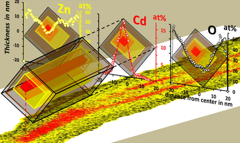
Quantitative 3D reconstruction of the structure and composition of a CdTe quantum box in a ZnTe nanowire. Rueda-Fonseca et al., Nano Letters, 2016
The NANO3D project: This project aims at developing a fast and easy method for the quantitative 3D reconstruction of core-shell nanostructures. The method requires a maximum of two X-ray maps acquired at two different tilt angles, preferably perpendicular to each other. The method is based on the modelling of the nanostructure cross-section using a series of imbricated ellipses whose dimensions are defined by their major and minor diameters. The number of ellipses depends on the number of chemical phases which are identified from the concentration profiles determined with the IZAC code. The position and orientation of each ellipse are determined by the coordinates of their respective centers and the overall tilt of the nanostructure, respectively. More sophisticated models, using hexagons or rectangles instead of ellipses, have been developed to consider the facetted sidewalls of crystalline nanostructures. These models are based on the elliptical model, by constructing the tangents to an ellipse, and hence, are defined by the same parameters, which is useful when comparing models. The method was applied for reconstructing core-shell nanostructures on (Mg, Mn, Cd, Zn)(Te,Se) nanowires, (Al, Cu, Sn)(Si,Ge) nanowires, (In,Ga)As nanowires and (Pt, Co) nanoparticles.